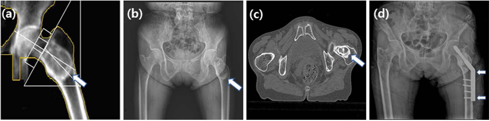
Fig. 1. In December 2010, a lesion (arrow) in the proximal femur (a) was observed by BMD. In February 2011, a lesion (arrow) in the proximal femur was observed using the pelvis AP image (b). In March 2011, a benign bone tumor, such as liposclerosing myxofibrous, was observed in the proximal femur, intertrochanteric region in both hip CT (c). In April 2011, an intramedually nail (arrows) in the left femur neck and shaft was observed using the pelvis AP image (d).