Splenic Absorption of Radiopharmaceutical in Systemic Bone Scans Performed after Liver Transplantation

Case Report
Sang-Hyeong Kil*Kyung-Nam JoYung-Hyun Lim

Abstract

A technetium-labeled phosphate bone scan was found to detect bone fractures and bone metastasis in the early stages compared to general radiographs. Therefore, bone scans are among the most frequently performed nuclear medical imaging examinations. However, non-osseous radiopharmaceutical uptake on bone scans is unusual. Herein, we report a case of diffuse splenic absorption of Tc-99m dicarboxypropane diphosphonate in patients who underwent liver transplantation.

Keyword


INTRODUCTION

Bone scans using technetium-labeled phosphate were found to detect bone fractures and metastasis in the early stages compared to general radiographs [1]. Therefore, bone scan is a commonly used modality for the evaluation of musculoskeletal lesions [2]. However, non-osseous radiopharmaceutical uptake on bone scans is often unexpected. Here, we report a case of diffuse splenic uptake of Tc-99m dicarboxypropane diphosphonate (DPD), which resembles a nuclear medicine spleen scan.

CASE PRESENTATION

A 47-year-old female presented with a history of alcoholic liver cirrhosis and splenomegaly underwent liver transplantation. The patient complain of lower backache and whole skeletal pain; a bone scan was performed to evaluate generalized osseous pain. A after 3h of intravenous injection oh 925 MBq Tc-99m DPD, whole-body images were recorded using a low-energy high-resolution collimator with a dual-head gamma camera (Symbia E, Siemens, USA)(Fig. 1).

http://dam.zipot.com:8080/sites/KJNMT/images/KJNMT-23-003_image/Figure_kjnmt_27_01_03_F1.png

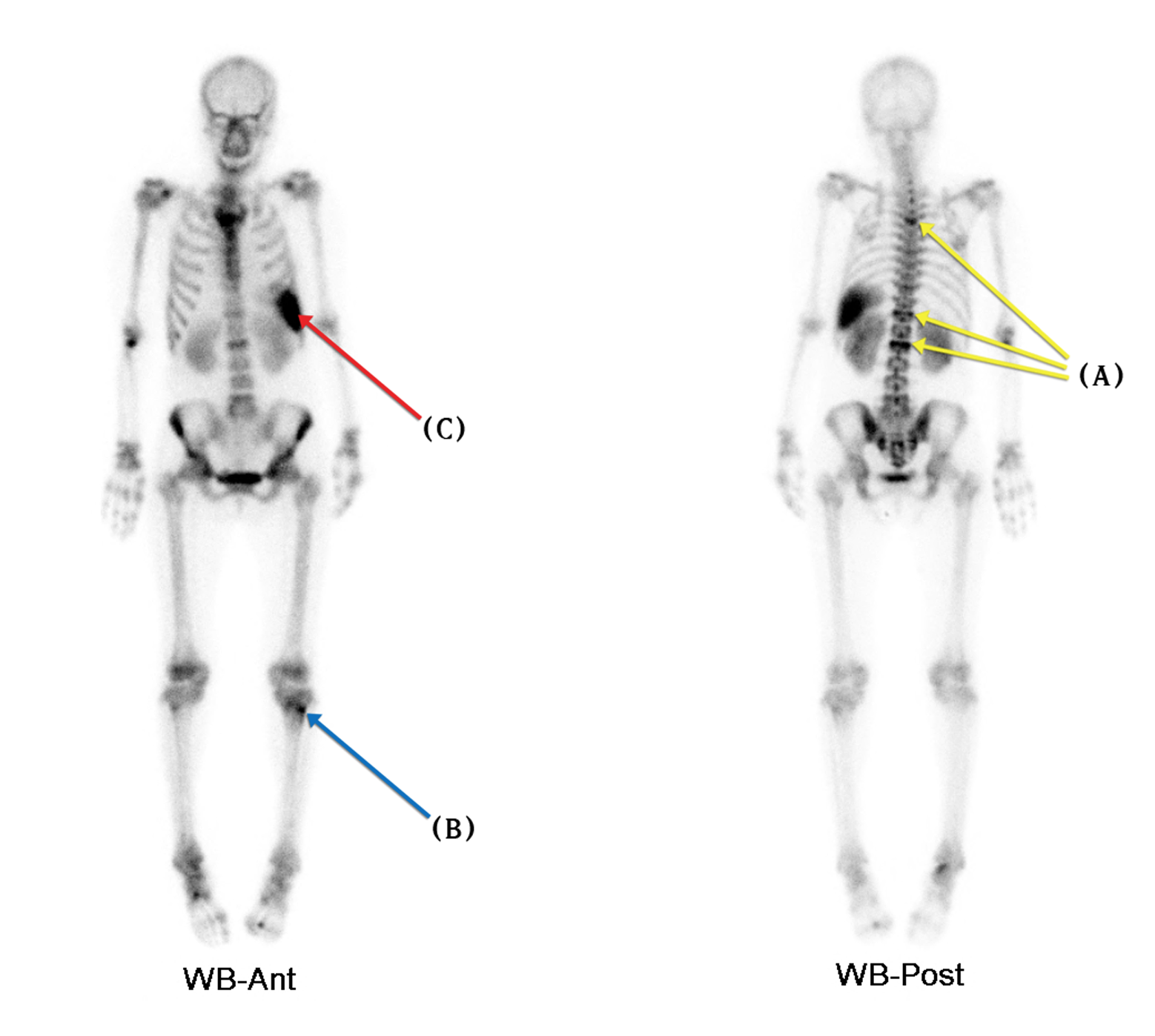
Fig. 1. Bone scan images show increased abnormal osseous uptake (A) on T4, T12, and L2 levels (compression fractures) and (B) left proximal tibia (suggestive traumatic bone lesion). Furthermore, they show increased (C) diffuse splenic uptake of radiopharmaceutical resembling nuclear medicine spleen scan.

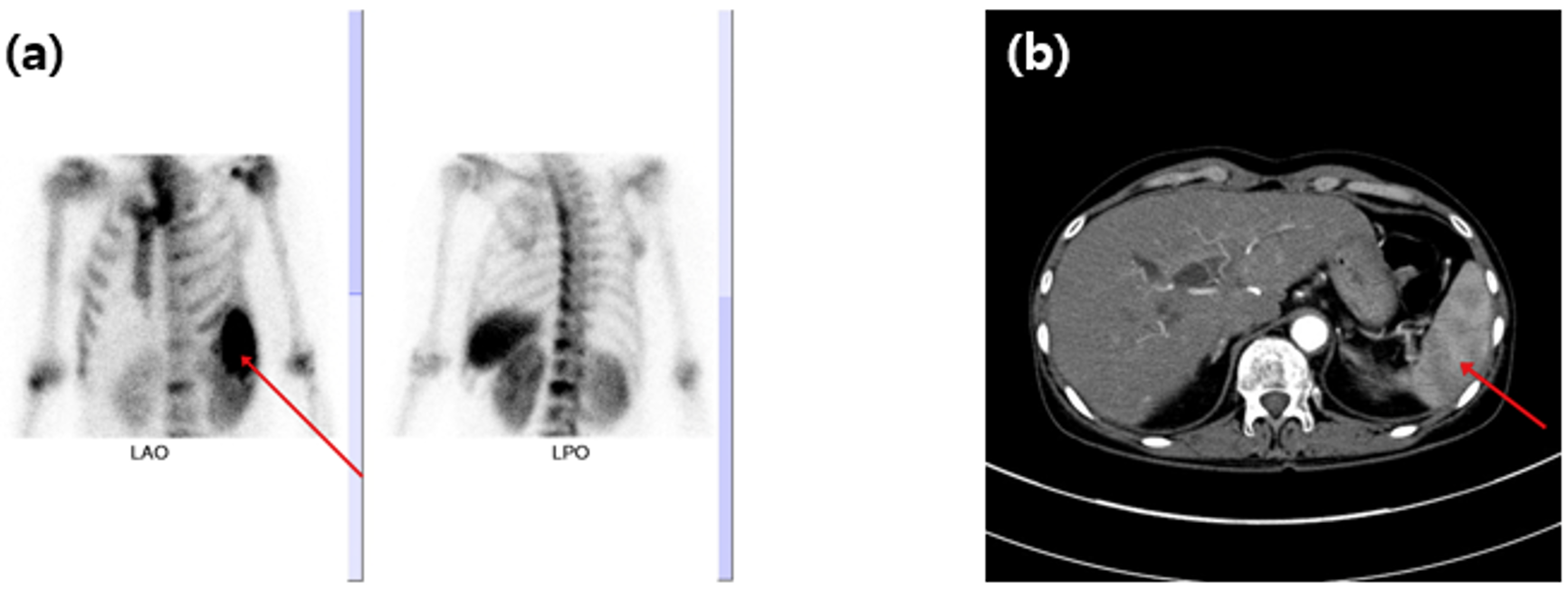
Bone scan images showed an increased diffuse splenic absorption of Tc-99m DPD (Fig. 2a). Contrast-enhanced computed tomography (CT) was performed. In this study, we performed contrast-enhanced CT, which revealed multiple poorly marginated, hypo-enhancing splenic lesions corresponding to infarction in the arterial and portal venous phases (Fig. 2b).

http://dam.zipot.com:8080/sites/KJNMT/images/KJNMT-23-001_image/Figure_kjnmt_27_01_03_F2.png

Fig. 2. Diffuse splenic uptake Tc-99 m DPD bone scan imaging (a) and contrast-enhanced CT imaging (b) findings were consistent with splenic infarction. Correlative contrast-enhanced computed tomography images are located on the bone scan images, with spleen infarction lesions marked by an arrow.

DISCUSSION

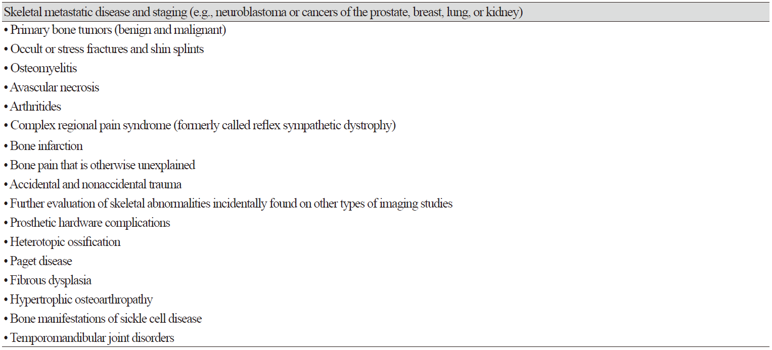
Bone scan images showed increased radiopharmaceutical absorption in lesions with increased osteoblastic activity. Table 1 lists common clinical indications for bone scans [1].

Table 1. Common Clinical Indications

http://dam.zipot.com:8080/sites/KJNMT/images/KJNMT-23-001_image/Table_kjnmt_27_01_03_T1.png

Unexpected radiopharmaceutical uptake in the spleen is occasionally observed during bone scans. The mechanism of splenic uptake of bone-seeking radiopharmaceuticals has been associated with the following finding [3-12]: residual radioactivity from a previous colloid scan, misadministration of radiocolloid, excessive aluminum ions from the generator, excessive serum aluminum, injection of radioiodinated contrast medium following bone agent injection, unknown metastatic calcification, hepatic necrosis, conditions of iron therapy, and compartmental sequestration. In this case, we can explain as splenic uptake of Tc-99m DPD was caused by multiple poorly marginated hypo-enhancing splenic lesions corresponding to the infarction.

References

1 Bartel TB, Kuruva M, Gnanasegaran G, Beheshti M, Cohen E-J, Weissman A-F, et al. SNMMI procedure standard for bone scintigraphy 4.0. J Med Radiat Sci. 2018;46(4):398-404 

2 Sanders TG, Parsons III TW. Radiographic imaging of musculoskeletal neoplasia. Cancer Control 8.3. 2001;8(3):221-31.   

3 Kalimuthu LM, Nazar AH, Pradhan PK. Idiopathic hepatic and splenic uptake of 99mTc-methylene diphosphonate. Indian J Nucl Med. 2020;35(3):238-40. 

4 Zuckier LS, Leonard MF. Nonosseous, nonurologic uptake on bone scintigraphy: atlas and analysis. Semin Nucl Med. 2010;40(4):242-56.   

5 Pierre DM, Annegret L, Julien M, Luc-Matthieu F, Izzie Jacques N. Spleen Uptake on Bone Scan After Frequent Platelet and RBC Transfusions. Clin Nucl Med. 2016;41(10):802-3.   

6 Shukla LW, Lin DS, Kutka N. Splenic uptake in bone imaging. Semin Nucl Med. 1988;18(1):71-3.   

7 Koizumi M, Suzuki T, Takahashi S, Ogata E. Transient splenic accumulation of Tc-99m HMDP caused by megaloblastic anemia. Clin Nucl Med. 2000;25(12):1024-7.   

8 Verma S, Kumar N, Kheruka SC, Gambhir S. Extraosseous 99mTc-methylene diphosphonate uptake on bone scan: Unusual scenario. Indian J Nucl Med. 2016;31(4):280-2.   

9 Joanie M. Idiopathic hepatic uptake of 99mTc methylene diphosphonate: A case report. J Nucl Med Technol. 2001;29(1):32-6. 

10 Hung JC, Ponto JA, Hammes RJ. Radiopharmaceutical-related pitfalls and artifacts. Semin Nucl Med. 1996;26(4):208–55. 

11 Zhang W, Chen B, Deng H, Yang T, Ou X. Hepatic and splenic uptake on bone scintigraphy in patients with intravenous administration of 99mTc methylene diphosphonate prior to gadolinium-containing contrast. Clin Nucl Med. 2013;38(3):219-20.   

12 Shih WJ, Coupal J. Diffuse and intense Tc-99m HMDP localization in the liver due to hypoxia secondary to respiratory failure. Clin Nucl Med. 1994;19(2):116-20.