Abstract
A 78-year-old woman referred by the Department of Obstetrics and Gynecology was recommended for orthopedic surgery because a cystic lesion in the left femoral proximal area was found by bone mineral densitometry (BMD). Computed tomography (CT) scan and general X-ray performed in the orthopedic department found a benign tumor. An intramedullary nail was inserted. Curettage and bone graft were performed. A radiologic technologist is important for the morphological observation of the femur in femoral BMD.
Figures & Tables
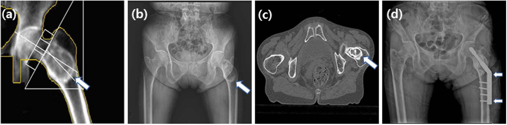
Fig. 1. In December 2010, a lesion (arrow) in the proximal femur (a) was observed by BMD. In February 2011, a lesion (arrow) in the proximal femur was observed using the pelvis AP image (b). In March 2011, a benign bone tumor, such as liposclerosing myxofibrous, was observed in the proximal femur, intertrochanteric region in both hip CT (c). In April 2011, an intramedually nail (arrows) in the left femur neck and shaft was observed using the pelvis AP image (d).