Abstract
Bone densitometry is a disease in which bones are easily broken due to metabolic bone disease, and DXA is used as a clinical standard test. Although DXA is a good method with good accuracy and reproducibility, it is frequently subject to test errors in testing and result analysis and analysis. Therefore, it is important to recognize the error issues that radiologists should basically be aware of when performing bone density tests, prevent erroneous diagnoses and treatments resulting from the results, and reduce the unnecessary costs associated with them. aim. The inspection must be carried out if the quality control of the equipment is basically continuously performed well before the inspection. Before starting the examination, the patient's age, sex, race, weight, pregnancy status, and any foreign objects that can be removed should be checked, and the examination should be performed in the correct posture. In addition, it is important to analyze results consistently. Radiologists, who play the most important role in ensuring accurate examinations, need to be aware of the potential for errors in advance and develop the ability to deal with the potential for errors in each examination. For that reason, regular education is considered essential.
Figures & Tables
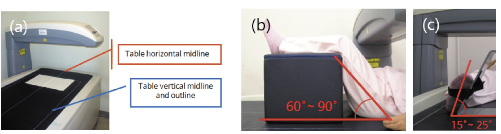
Fig. 1. The center line and outline of the examination table to maintain a consistent patient posture in bone mineral densitometry (a). During the lumbar examination, have the patient lie down in the middle in a comfortable state, and pull the legs slightly downward to avoid straining the spine in a relaxed state. Then, using the position block, bend your legs up to 60°-90° to maintain the posture by straightening the curved spine (b). In the femoral examination, the leg is rotated within 15°-25° with the leg relaxed, and the cervical axis of the femur is positioned so that it is parallel to the plane of the scan table (c).