Abstract
A 50-year-old female patient referred by the Department of Breast and Thyroid Surgery was recommended for orthopedic surgery because a lesion-like herniation pit was found in the left proximal femur in bone mineral densitometry (BMD). She was later diagnosed with bone metastasis based on a biopsy in orthopedic surgery. Pelvic X-ray and left thigh MRI were performed. An intramedullary nail was later inserted. The BMD is a diagnostic method based on numerical values, so it is inevitable that the bone shape may be neglected. This case report illustrates the importance of the radiotechnologist's observation of the bone shape. In our patient, it led to beneficial treatment.
Figures & Tables
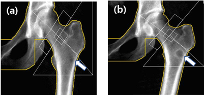
Fig. 1. A lesion-like herniation pit (arrow) was observed in the proximal femur in bone mineral densitometry (a) conducted in January 2017. Bone density imaging (b) performed again in January 2019 shows the same lesion (arrow) has enlarged.