The Pitfalls Medical Radiological Technologists Should Consider in Bone Densitometry

Review article
김 호성  Ho-Sung Kim*

Abstract

Bone densitometry is a disease in which bones are easily broken due to metabolic bone disease, and DXA is used as a clinical standard test. Although DXA is a good method with good accuracy and reproducibility, it is frequently subject to test errors in testing and result analysis and analysis. Therefore, it is important to recognize the error issues that radiologists should basically be aware of when performing bone density tests, prevent erroneous diagnoses and treatments resulting from the results, and reduce the unnecessary costs associated with them. aim. The inspection must be carried out if the quality control of the equipment is basically continuously performed well before the inspection. Before starting the examination, the patient's age, sex, race, weight, pregnancy status, and any foreign objects that can be removed should be checked, and the examination should be performed in the correct posture. In addition, it is important to analyze results consistently. Radiologists, who play the most important role in ensuring accurate examinations, need to be aware of the potential for errors in advance and develop the ability to deal with the potential for errors in each examination. For that reason, regular education is considered essential.

Keyword


서론

골다공증은 골량의 감소와 미세구조의 이상을 특징으로 하는 대사성 골 질환으로 뼈가 약해져 쉽게 부러지는 상태의 질병으로[1], 임상에서 골다공증을 진단하는 데 널리 사용되는 골밀도(Bone Mineral Density, BMD) 측정은 일반적으로 골절 위험의 대용으로 사용된다(2). 이러한 특징의 골다공증 진단에 사용되고 있는 이중 에너지 X선 흡수법(Dual-energy X-ray Absorptiometry, DXA)은 허용 가능한 정밀도 오차, 우수한 정밀도 및 재현성으로 BMD를 측정하는 기준 방법이다[3]. 하지만 DXA 검사의 오류는 영상의 획득 및 결과분석에서 일반적으로 보고된 것처럼 자주 발생한다[4-11]. 따라서 다른 많은 진단 검사와 마찬가지로 DXA 스캔은 BMD 측정에 영향을 미칠 수 있는 이상 여부를 해석하는 의사와 방사선사가 비판적으로 평가해야 한다[3]. 골밀도 영상 보고의 오류는 1% 미만에서 30% 이상까지 다양한 비율로 잘 알려져 있다[12-14]. 이는 임상에서 BMD 결과에 영향을 미칠 수 있는 다양한 인공물 및 질병 과정에서 발생하는 다양한 문제인식은 DXA 스캔의 최적 해석에서 매우 중요할 수 있다[15]. 최근 연구는 국제임상골밀도학회(International Society for Clinical Densitometry, ISCD)의 약 6,000명의 회원을 대상으로 DXA 보고서의 품질에 대한 인식을 조사했습니다[6]. 조사를 받은 임상의의 21% (3,488명 중 743명)와 기술자의 32% (2,362명 중 754명), 대부분의 임상의(71%)와 수많은 방사선사(45%)가 잘못된 DXA 해석을 한 달에 한 번 이상 보고 있다고 보고했다. 거의 모든 임상의(98%)가 질이 낮은 DXA 보고서는 환자 관리에 유해하다고 느꼈으며, 또한 DXA 스캔 전에 인구통계 정보(연령, 성별 등)가 잘못 입력되었기 때문에 오해를 초래하는 보고서나 부정확한 보고서가 생성될 수 있음이 발견되었다. 따라서 방사선사가 골밀도 검사를 수행함에 있어 기본적으로 알아야 할 오류 문제를 인식하고 결과에 따라 발생할 수 있는 잘못된 진단과 치료를 방지하고, 그에 따라 발생하는 불필요한 비용을 줄이기 위함을 목적으로 한다.

본론

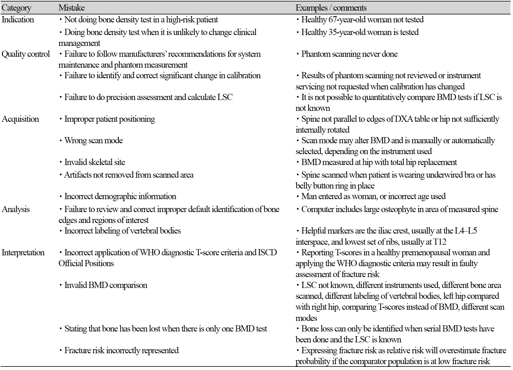
DXA 골밀도 검사 오류의 주요 원인은 운영자의 관리 하에 있다[15, 16]. 고품질 DXA 보고서를 생성하려면 장비 교정 변경, 부적절한 환자 위치 결정 또는 분석, 교락 아티팩트 인식, T 및 Z 점수 계산을 위한 참조 데이터베이스의 올바른 선택 등 오류 잠재력 원인을 이해해야 한다[9]. 아래의 도표(Table. 1)는 흔히 발생하는 골밀도 검사의 실수를 나타낸 것이다[17]. 따라서 아래의 내용을 기준으로 방사선사의 검사 및 결과분석에 대한 기술적 요인으로 발생할 수 있는 오류의 원인을 분석하고 대처방안을 기술하려 한다.

Table 1. Programs in STADEN package

http://dam.zipot.com:8080/sites/KJNMT/images/KJNMT-23-009_image/Table_kjnmt_27_01_05_T1.png

1. 검사 전 확인

1. 1. 나이, 성별, 인종, 체중

골밀도 검사 후 진단에서 가장 기본적으로 확인해야 하는 것은 환자의 정확한 신원 정보이다. 또한 인구통계에는 환자의 이름, 생년월일, 성별, 병원등록번호 또는 기타 식별자, 신장, 체중, 스캔일, 보고일, 추천 의사의 이름, 판독의의 이름 및 BMD 시설의 이름과 위치를 포함해야 한다[6, 18-21]. 체중과 키는 BMD 시설에서 측정해야 한다[22, 23]. 측정을 수행할 수 없는 예외적인 상황(예: 환자가 서있을 수 없는 경우)을 제외하고는 환자가 보고한 값이나 다른 의료 종사자가 제공한 측정치를 사용하지 말아야 하며 키 또는 체중 데이터가 BMD 시설에서 직접 측정되지 않은 경우는 보고서에 표시해야 한다[24]. 골무기질(Bone Mineral Content, BMC), BMD 및 골 면적의 측정치는 조직 구성의 영향을 받는다[25]. Laskey 등은 전신 스캔 모드와 척추 스캔 모드의 BMC, 골 면적 및 BMD 결과를 비교하여 전신 스캔 모드가 10 ㎝ 이상의 깊이에서 BMC 및 골 면적의 값이 높게 나타났으며, 깊이 또〮〮는 두께가 감소하면 BMD가 증가했다, 다만 팬텀과 물을 이용하여 연구가 진행되어 실제 생체 내 상황과는 다를 수 있지만 비만인 경우 BMD 정확도가 떨어질 수 있다고 보고하였다[26]. 그리고 Svendsen 등은 Van Loan과 Mayclin [27]이 수행 한 것처럼 체중과 조성의 큰 변화가 DXA의 뼈 가장자리를 정확하게 감지하는 능력에 영향을 미치고 뼈의 면적 추정을 변경할 수 있다는 것을 제안했다. 또한 Gary R. Hunter et al. 의 연구에서는 적당한 체중은 BMD 손실과 관련이 없고, 저항 훈련 및/또는 점프 훈련과 같은 고강도 훈련은 유지 관리 또는 체중 감량 중 BMD 증가와 관련이 있고, 체중 감량 중 칼슘 및 비타민 D 섭취 유지는 BMD 유지와 관련이 있다고 보고하였다[28].

1. 2. 임신여부

임신의 경우 태아의 방사선 피폭의 우려로 검사가 불필요하다고 판단하는 사람들과의 법적인 소송을 피하기 위해서는 특별한 이유가 없는 경우를 제외하고는 검사를 진행하지 않는 것이 바람직하다. M Kaur et al.의 연구에서 피크 골량에 영향을 미치고 후속 삶에서 골다공증을 일으킬 위험에 영향을 줄 수 있는 일반적인 생리학적 사건으로, 46명의 정상 여성의 임신 전과 출산 직후 이중 에너지 X 선 흡수 방법에 의해 BMD를 측정하고, 임신 실패 30명의 대조 여성과 비교한 연구에서 임신한 여성과 임신하지 않은 여성 사이에는 차이가 없다고 보고하였다[29]. 그리고 단일 광자 흡수(Single Photon Absorptiometry, SPA) 및 이중 광자 흡수(Dual Photon Absorptiometry, DPA)에 의해 결정된 피질 또는 뼈의 골밀도에 유의한 변화는 보이지 않았다는 보고도 있다[30-33]. 하지만, Naylor et al.은 임신 중 피질 부위의 BMD 증가와 소주골 부위의 BMD 감소를 보고했고[34], 요추 BMD는 임신 중 3.3% 감소하는 것으로 보고되었다[35]. 따라서 임신의 경우 객관적인 임상자료 증거를 기준으로 태아의 피폭 우려에 대하여 검사 시기를 미루는 등의 조정으로 대처해야 한다.

2. 장비 관리

2. 1. Daily QC & Periodic QC

BMD 측정의 일관성을 유지하기 위해서는 BMD 측정의 QC (Quality Control)가 필수적이다[36-38]. DXA를 이용한 BMD 측정의 QC는 정밀도와 정밀도의 오차 범위를 최소화하기 위해 수행된다. 이를 위해 환자의 상태, 측정 장비 및 방사선사의 경험으로 인한 오차 범위는 품질 관리에 의해 최소화되어야 한다[39]. 따라서 ISCD에서 권고하는 내용에 부합하여 장비 정도관리와 검사자(방사선사) 정도관리가 기본적으로 수행되어야 한다.

3. 이물질 유무

골다공증 검사 시 나타날 수 있는 이물질의 유형은 제게 가능한 것과 그렇지 않은 것으로 구분할 수 있다. 액세서리는 검사 전 제거 할 수 있도록 접수 단계에서 설명하고, 그 외 수술에 의한 외과적 고정 장치나 검사를 위한 조영제 및 칼슘제제 약물복용 등에 관한 사항을 철저하게 스크리닝 해 주어야 한다. DXA를 사용한 최적의 골밀도 측정은 아티팩트를 최소화해야 한다[40]. 이들 중 대부분이 내부에 있으며 외과적 고정 장치, 골관절염, 석회화 된 대동맥 또는 임플란트 등을 제거 할 수 없다[41-47]. 또한 위장관에서 제거되지 않은 바륨과 테크네튬은 DXA 측정 결과에 악영향을 미친다[24, 48]. 칼슘 정제 섭취도 유사한 영향을 미치므로 척추 DXA 결과에 혼란을 줄 것이라고 추정된다. 칼슘 정제는 피할 수 있는 아티팩트일 수 있으므로 많은 DXA 센터와 ACR (American College of Radiology) / RSNA (Radiological Society of North America)는 일상적으로 스캔 수행 24시간 전에 칼슘 정제를 섭취하지 말라고 조언한다[49]. 흥미롭게도, DXA 스캐닝 전에 칼슘 정제를 피하라는 이러한 권장 사항은 다른 약물이나 보충제로 확장되지 않으며 추상적 형태로 제시된 생체 외 데이터 결과는 많은 정제가 팬텀 BMD 측정을 변경할 수 있음을 시사한다[50].

4. 골밀도 검사

올바른 검사를 위해 가장 기본은 환자의 올바른 자세 잡이다. 자세 잡이를 일관적으로 유지하기 위해 요추 검사 시 검사 테이블에 그려져 있는 중앙선과 외곽선을 이용하여(Fig. 1-A) 편안한 상태로 가운데에 눕게 하고 힘을 뺀 상태에서 척추에 무리가 가지 않도록 다리를 아래쪽으로 살짝 당기고, 포지션 블럭을 이용하여 60°-90° 정도로 다리를 구부려 올려 굴곡이 되어 있는 척추를 펴서 자세를 유지하여 움직임이 없도록 하고(Fig. 1-B), 대퇴부 검사에서는 다리에 힘을 뺀 상태에서 15°-25° 내 회전을 시키고 대퇴골의 경부 축이 스캔 테이블의 평면과 평행이 되도록 위치하는 것이 중요하다(Fig. 1-C). 다만 여러 사람이 근무하는 경우 항상 똑같은 방법으로 환자 자세를 유지하는 것이 방사선사 간 정밀도 오차를 줄이는데 도움이 될 것이다. 또한 검사는 제조업체 프로토콜 준수하고, 적절한 위치 지정 및 하위 영역 할당, 뼈 추적, 관심 영역 결정 및 품질 보증을 포함하여 스캐닝 수행 방법의 모든 기술적 측면에서 주의를 기울여야 한다[6, 51-62].

http://dam.zipot.com:8080/sites/KJNMT/images/KJNMT-23-009_image/Figure_kjnmt_27_01_05_F1.png

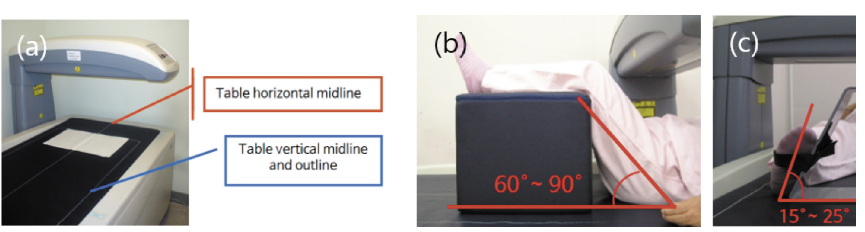
Fig. 1. The center line and outline of the examination table to maintain a consistent patient posture in bone mineral densitometry (a). During the lumbar examination, have the patient lie down in the middle in a comfortable state, and pull the legs slightly downward to avoid straining the spine in a relaxed state. Then, using the position block, bend your legs up to 60°-90° to maintain the posture by straightening the curved spine (b). In the femoral examination, the leg is rotated within 15°-25° with the leg relaxed, and the cervical axis of the femur is positioned so that it is parallel to the plane of the scan table (c).

4. 1. Spine

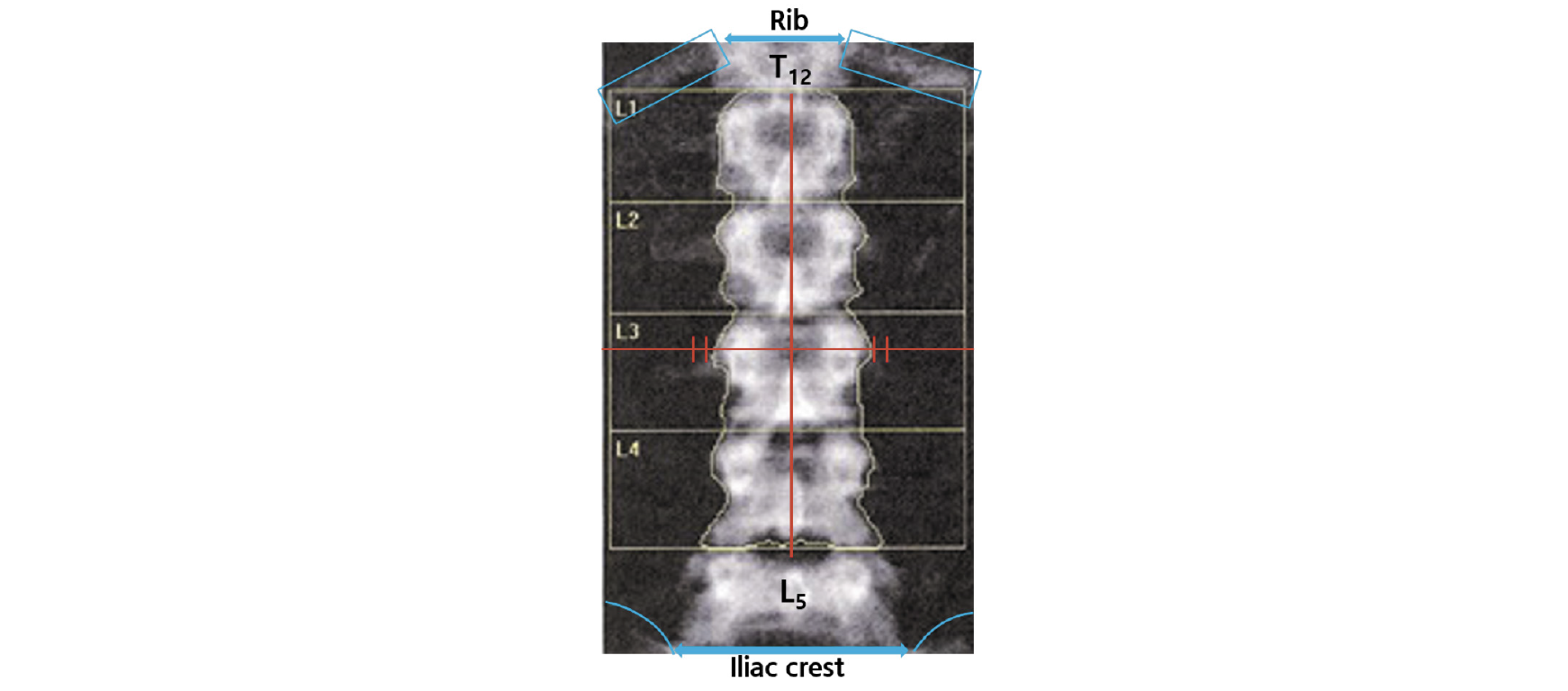
L1-L4 BMD 평가에서 극돌기는 곧은 정중선의 중심에 있어야 하며 천골(장골)의 일부와 갈비뼈가 있는 척추의 일부(일반적으로 T12)를 포함해야 하며, 척추의 회전이 없어야 한다. 만약 정중선을 기준으로 방향에 상관없이 60°까지는 BMD가 거의 20% 가까이 감소한다[63]. 이는 BMD가 영역별 BMC로 평가되기 때문이다. 따라서 척추 측만증 환자의 경우 정확한 골밀도 측정이 어려울 수 있다. 그리고 척추의 형태학적인 식별은 천골부터 번호가 매겨져야 6개의 요추 또는 변동성이 있는 12번 늑골로 인한 잘못된 척추 식별 오류를 방지할 수 있다. 그리고 L1은 U자형, L2는 V자형, L3은 Y자형, L4는 H자형이고 L5는 대문자 I가 옆으로 누운 형태로 나타난다(Fig. 2). 또한 측면 보기에서 12번째 늑골은 L1과 겹치고 골반은 L4와 겹치므로 라벨링이 어려울 경우 요추를 식별하는 데 도움이 될 수 있다[64].
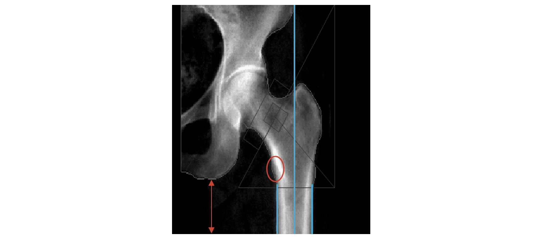
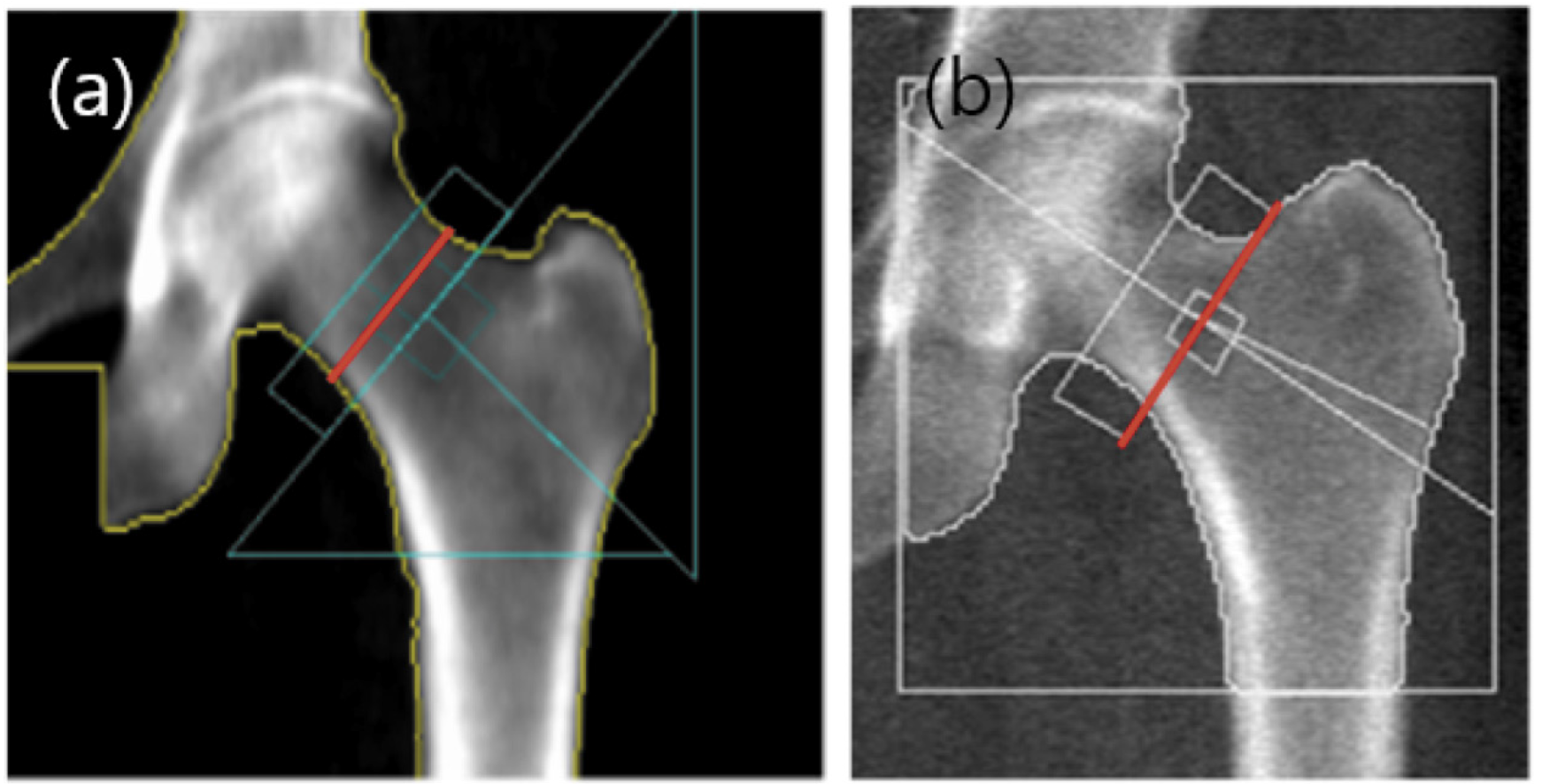
http://dam.zipot.com:8080/sites/KJNMT/images/KJNMT-23-009_image/Figure_kjnmt_27_01_05_F2.png

Fig. 2. In the L1-L4 BMD assessment, the spinous process should be centered on a straight midline and should include part of the sacrum (ilium) and part of the spine with the ribs (usually T12), and there should be no rotation of the spine.

4. 2. Femur

대퇴골 경부의 BMD 평가에서 해부학적인 자세는 소전자가 살짝 보일 정도의 자세이다. 샤프트는 일직선의 되어야 하고 소프트 티슈가 충분하게 포함되어야 한다(Fig. 3). 만약 소전자가 보이지 않는 경우는 골밀도가 증가하는 경향을 보인다. Femoral neck box는 GE lunar와 Hologic의 분석에서 차이가 있다. GE lunar의 경우는 대퇴골두와 전자의 중간 지점인 가장 좁은 부분에 위치하며(Fig. 4-A), Hologic의 경우는 대퇴골 경부의 가장 말단부에 위치한다(Fig 4-B)[65]. 근위 대퇴골의 경우 왼쪽을 측정할 수 없거나 유효하지 않은 경우 또는 이전에 오른쪽 엉덩이를 측정한 적이 있는 경우가 아니면 왼쪽을 측정해야 한다[66, 67].

http://dam.zipot.com:8080/sites/KJNMT/images/KJNMT-23-009_image/Figure_kjnmt_27_01_05_F3.png

Fig. 3. In the BMD evaluation of the femoral neck, the anatomical position is such that the lesser trochanter is slightly visible. The shaft should be straight and contain plenty of soft tissue.

http://dam.zipot.com:8080/sites/KJNMT/images/KJNMT-23-009_image/Figure_kjnmt_27_01_05_F4.png

Fig. 4a, 4b. The emoral neck box was different in the analysis of GE lunar and Hologic. In the case of GE lunar, it is located at the narrowest part midway between the femoral head and the former (a), and in the case of Hologic, it is located at the most distal part of the femoral neck (65) (b).

5. 결과분석

최소 2개의 골격 부위를 스캔하고 보고해야 한다[18, 51, 58, 68-71]. 그리고 유효한 스캔이 있는 각 골격 부위에 대해 보고된 골밀도 결과는 절대 BMD (g/㎠에서 소수점 이하 3자리까지) 및 T-점수(소수점 1자리까지)가 포함되어야 한다[18, 51, 71, 73].

5. 1. Spine

요추의 경우 보고서에 결과의 그래픽 표현이 포함된 경우 그래프에는 해석에 실제로 사용된 척추에 대한 데이터 및 참조 곡선이 표시되어야 한다[18]. 해당 척추의 T-점수가 다음으로 높은 값을 갖는 척추의 T-점수보다 1 표준 편차 이상 큰 경우 특정 척추를 제외하는 것을 고려할 수 있다[18, 51, 74]. 밀도가 높은 척추뼈를 반드시 배제해야 하는 것은 아니지만, 인공물의 원인을 평가하고 척추 분석에서 포함 여부를 결정해야 한다.

5. 2. Femur

근위대퇴골의 경우 전체 고관절, 대퇴골 경부 및 전자 영역에 대한 결과를 보고해야 한다[73-77]. 검사가 종료된 후 반드시 영상을 확인하고 air scan이 있는지의 여부를 살피는 것 또한 중요하다(Fig. 4). 만약 아래와 같은 영상이 확인되면 마른 체형인지를 확인하고 적정위치에서 검사가 이루어졌는지 확인하고 rice bag이나 스캔모드를 변경해서 재검사를 진행하고 재검사시 동일한 조건으로 검사를 수행할 수 있도록 메모를 해두어야 한다.

또한 원위 대퇴골 검사는 대퇴골의 회전에 따라 골 면적에 차이가 발생하여 결과분석에 영향이 있다. 즉 골면적이 증가하면 골밀도가 감소하고, 골면적이 감소하면 골밀도가 증가한다. 따라서 골밀도 검사 시설에서는 분석 시 각 장비회사의 프로토콜에 따라 자동분석과 수동 분석을 적절하게 병행하는 것이 검사의 정확도 측면에서 유리하다. 특히 재검의 경우는 ROI (Region of Interest) Copy 기능을 이용하여 이전 검사와의 동일한 영역으로 일관되게 비교 분석하는 것이 바람직하다(Fig. 7).

http://dam.zipot.com:8080/sites/KJNMT/images/KJNMT-23-009_image/Figure_kjnmt_27_01_05_F5.png

Fig. 5. After the femur examination, the air scan is confirmed as shown in the red circle in the result analysis.

http://dam.zipot.com:8080/sites/KJNMT/images/KJNMT-23-009_image/Figure_kjnmt_27_01_05_F6.png

Fig. 6. This is a picture of the change in bone area according to rotation. Area and BMD are inversely proportional.

http://dam.zipot.com:8080/sites/KJNMT/images/KJNMT-23-009_image/Figure_kjnmt_27_01_05_F7.png

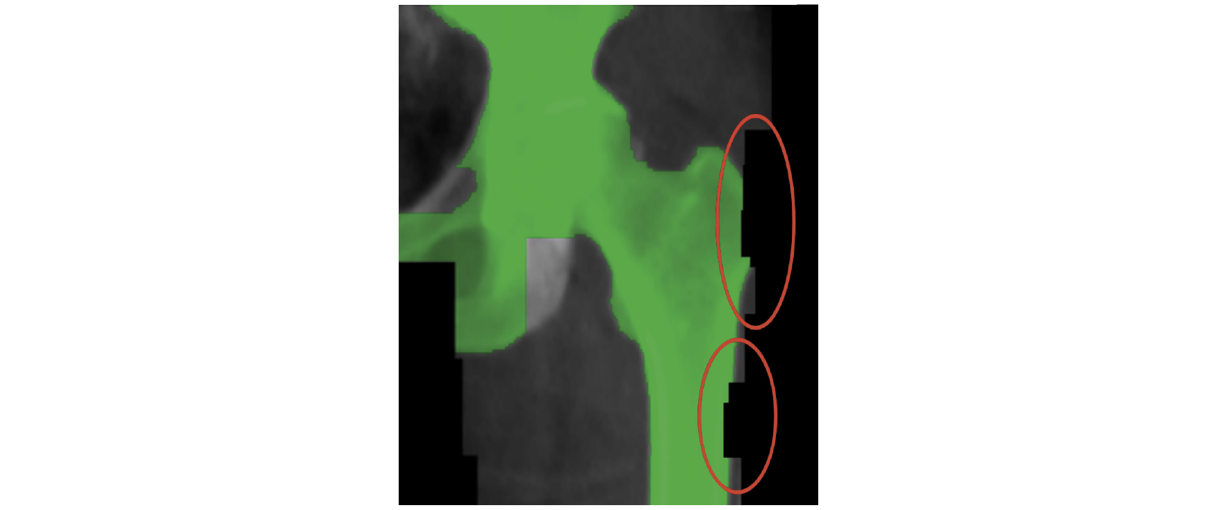
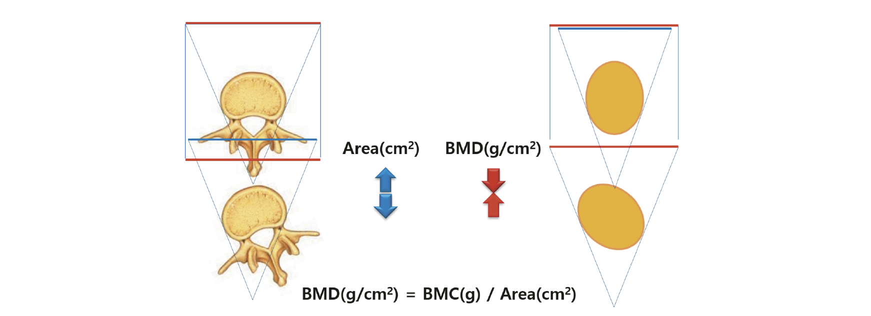
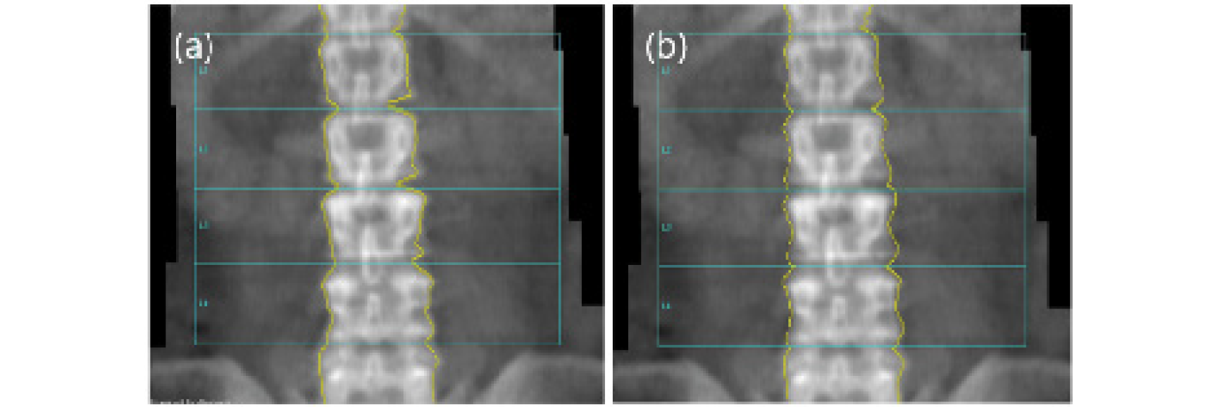
Fig. 7. This picture shows the difference between automatic analysis and manual analysis. If the edge of a bone cannot be recognized, a difference in BMD occurs when the edge is mapped according to the standards recommended by each manufacturer (a; auto mapping, L1~L4: BMD=0.857, T-Score=-2.4, b; manual mapping, L1~L4: BMD=0.788, T-Score=-2.7).

5. 3. 질병, 퇴행성 변화, 공간점유병소 등

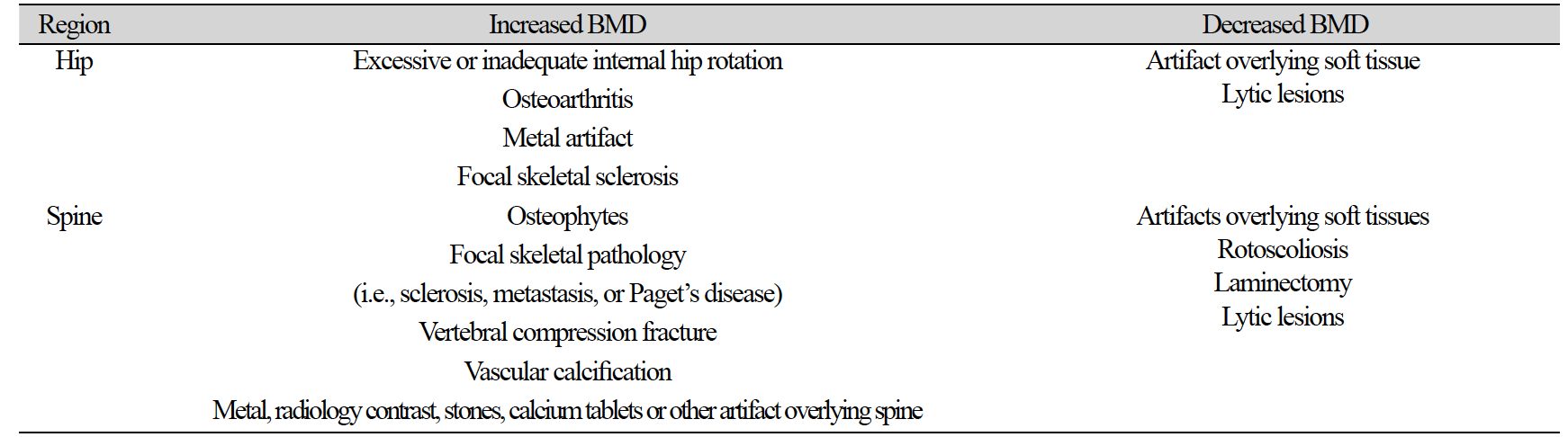
검사하는 부위의 구조적인 이상이나 인공물 등은 결과에 상당한 영향을 미칠 수 있다. 체중과 같이 BMD 결과를 감소시키거나 증가시킬 수 있는 BMD와는 독립적인 요인이 있다(Table 2)[19].

Table 2. Factors modifying bone mineral density (BMD)*

http://dam.zipot.com:8080/sites/KJNMT/images/KJNMT-23-009_image/Table_kjnmt_27_01_05_T2.png

*Extremes of body weight or significant change (more than 10%) in body weight can have unpredictable effects on BMD and affect serial measurements.

검사 전 이물질 제거가 되지 않은 경우는 ROI 설정에 오류가 발견되거나 분석에 영향이 있으므로 반드시 검사 영역에 포함 시켜서는 안 된다(Fig. 8).

http://dam.zipot.com:8080/sites/KJNMT/images/KJNMT-23-009_image/Figure_kjnmt_27_01_05_F8.png

Fig. 8. The picture on the left is a picture of the test performed without removing the urine line in the bone density test, and the picture on the right is a picture of the result analysis. What the arrows in the picture teach is that the automatic analysis is not recognizing the bones (a, b).

그리고 원위 대퇴골 검사의 경우는 Femur Neck 분석에서 ROI Box영역이 좌골과 겹치는 경우가 발생하는데 이에 따라 골밀도의 차이가 발생할 수 있기 때문에 주의해야 한다(Fig. 9).

http://dam.zipot.com:8080/sites/KJNMT/images/KJNMT-23-009_image/Figure_kjnmt_27_01_05_F9.png

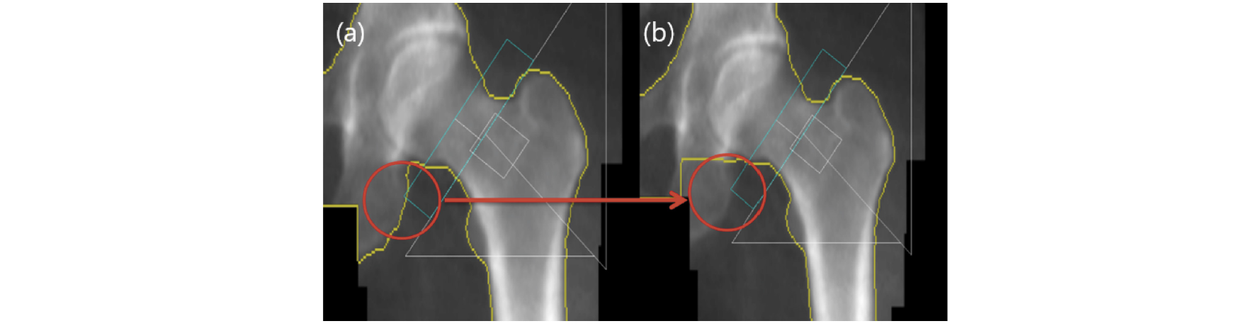
Fig. 9. In the analysis of Femur test results, if Ischium overlaps in the neck ROI box, a difference in BMD occurs. Therefore, the overlapping part must be removed by manual mapping (a; Ischium is overlapped with Femur neck ROI box (neck BMD: 1.111 g/cm2, Total: 1.166 g/cm2, b; Ischium removed from Femur neck ROI box (neck BMD: 1.136 g/cm2, Total: 1.189 g/cm2). The neck increased by 2.2% and the total increased by 2.0%.
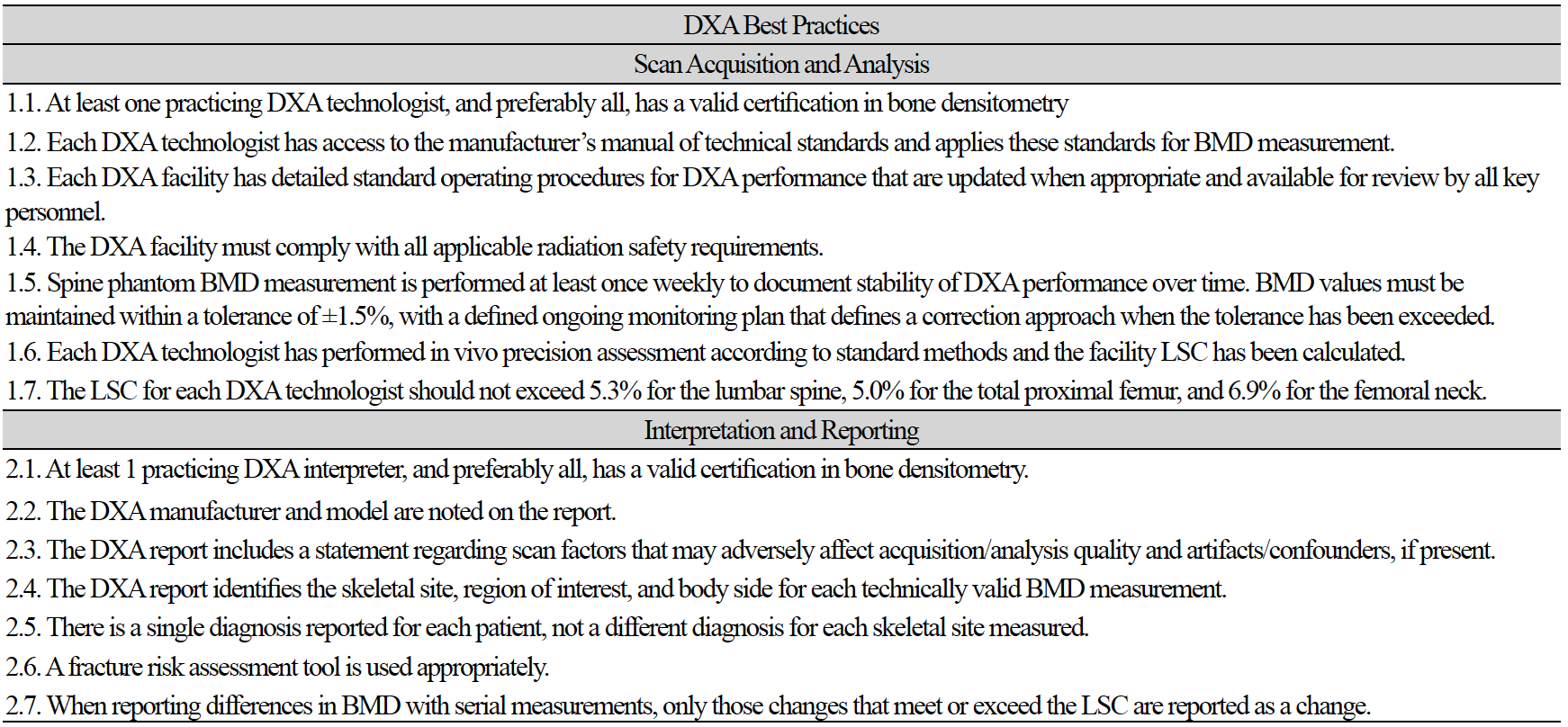
결론

골밀도 검사의 경우 여러 가지의 영향으로 인하여 오류 발생 가능성이 많다는 것은 본문에서 나열된 많은 연구를 통해 알 수 있다. 정확한 검사를 위해 가장 중요한 역할을 하는 방사선사는 오류 가능성에 대하여 미리 인식하고 있는 것이 중요하며, 검사마다의 오류 가능성에 대하여 준비 대처할 수 있는 능력을 키우는 것이 필요하다. 그러기 위해서는 정기적인 교육이 필수적이 요소라고 생각된다. 아래의 도표(Table 3)는 DXA 판독의사, 방사선사의 가이드 및 기대치를 제공하기 위한 것으로[9], 이러한 DXA 모범 사례를 준수하여 방사선사의 검사 및 결과분석에 대한 기술적 요인으로 발생할 수 있는 오류의 원인을 분석하고 대처하기 위한 기본적인 사항으로 각 병원에 특성에 맞게 적용하면 도움이 될 것으로 기대된다.

Table 3. Factors modifying bone mineral density (BMD)*

http://dam.zipot.com:8080/sites/KJNMT/images/KJNMT-23-009_image/Table_kjnmt_27_01_05_T3.png

BMD, bone mineral density; DXA, dual-energy X-ray absorptiometry; LSC, least significant change.

References

1 1. NIH consensus development panel on osteoporosis prevention, diagnosis, and therapy. Osteoporosis prevention, diagnosis, and therapy. JAMA 2001;285:785–95. 

2 2. Price RI, Walters MJ, Retallack RW, Henderson NK, Kerr D, Henzell S, et al. Impact of the analysis of a bone density reference range on determination of the T-score. Journal of Clinical Densitometry. 2003;6(1):51-62. 

3 3. Blake GM, Fogelman I. The role of DXA bone density scans in the diagnosis and treatment of osteoporosis. Postgraduate Medical Journal. 2007;83(982):509-17. 

4 4. Ruutiainen, Alexander T. Utilization and reporting of bone densitometry: what can the musculoskeletal radiologist do to help, rather than to hurt?. Academic Radiology. 2015;22(8):1030-3. 

5 5. Quint LE, Quint DJ, Myles JD. Frequency and spectrum of errors in final radiology reports generated with automatic speech recognition technology. Journal of the American College of Radiology, 2008;5(12):1196-9. 

6 6. Lewiecki EM, Binkley N, Petak SM. DXA quality matters. Journal of Clinical Densitometry. 2006;9(4):388-92. 

7 7. Gafni RI, Baron J. Overdiagnosis of osteoporosis in children due to misinterpretation of dual-energy x-ray absorptiometry (DEXA). The Journal of pediatrics. 2004;144(2):253-7. 

8 8. Johnston R, Colquhoun A, Wu W, Kim S. Quality assessment of bone density testing by DXA—Evaluation of technical and reporting deficiencies identified at a tertiary osteoporosis clinic. Journal of Clinical Densitometry. 2016;4(19):538-9. 

9 9. Lewiecki EM, Binkley N, Morgan SL, Shuhart CR, Camargos BM, Carey JJ, et al. Best practices for dual-energy X-ray absorptiometry measurement and reporting: International society for clinical densitometry guidance. Journal of clinical densitometry. 2016;19(2):127-40. 

10 10. Schousboe JT, Shepherd JA, Bilezikian JP, Baim S. Executive summary of the 2013 international society for clinical densitometry position development conference on bone densitometry. Journal of Clinical Densitometry. 2013;16(4):455-66. 

11 11. Çetin A, Özgüçlü E, Özçakar L, Akıncı A. Evaluation of the patient positioning during DXA measurements in daily clinical practice. Clinical rheumatology. 2008;27:713-5. 

12 12. Karahan AY, Kaya B, Kuran B, Altındag O, Yildirim P, Dogan SC, et al. Common mistakes in the dual-energy X-ray absorptiometry (DXA) in Turkey. A retrospective descriptive multicenter study. Acta Medica. 2017;59(4):117-23. 

13 13. Fenton JJ, Robbins JA, Amarnath ALD, Franks P. Osteoporosis overtreatment in a regional health care system. JAMA internal medicine. 2016;176(3):391-3. 

14 14. Promma S, Sritara C, Wipuchwongsakorn S, Chuamsaamarkkee K, Utamakul C, Chamroonrat W, et al. Errors in patient positioning for bone mineral density assessment by dual X-ray absorptiometry: effect of technologist retraining. Journal of Clinical Densitometry. 2018;21(2):252-9. 

15 15. El Maghraoui A, Roux C. DXA scanning in clinical practice. QJM: An international Journal of Medicine. 2008;101(8):605-17. 

16 16. Fuleihan GEH, Testa MA, Angell JE, Porrino N, Leboff MS. Reproducibility of DXA absorptiometry: a model for bone loss estimates. Journal of Bone and Mineral Research. 1995;10(7):1004-14. 

17 17. Lewiecki EM, Lane NE. Common mistakes in the clinical use of bone mineral density testing. Nature clinical practice Rheumatology. 2008;4(12):667-74. 

18 18. Siminoski K, Leslie WD, Frame H, Hodsman A, Josse RG, Khan A, et al. Recommendations for bone mineral density reporting in Canada. Journal-Canadian Association of Radiologists. 2005;56(3):178. 

19 19. Baim S, Binkley N, Bilezikian JP, Kendler DL, Hans DB, Lewiecki EM, et al. Official positions of the international society for clinical densitometry and executive summary of the 2007 ISCD position development conference. Journal of clinical densitometry. 2008;11(1):75-91. 

20 20. Stock JL, Waud CE, Coderre JA, Overdorf JH, Janikas JS, Heiniluoma KM, et al. Clinical reporting to primary care physicians leads to increased use and understanding of bone densitometry and affects the management of osteoporosis: a randomized trial. Annals of Internal Medicine. 1998;128(12_Part_1):996-9. 

21 21. Ridout R, Hawker GA. Use of bone densitometry by Ontario family physicians. Osteoporosis International. 2000;11:393-9. 

22 22. National Health and Nutrition Examination Survey III. Body measurement (anthropometry). 1988. Available at: www.cdc.gov. Accessed May 12, 2010. 

23 23. Lipman TH, Greisinger AJ, Sanders LL, Boyle MD, Shuster JJ, Johnson JV, et al. Evaluation of the accuracy of height assessment of premenopausal and menopausal women. Journal of Obstetric, Gynecologic & Neonatal Nursing. 2006;35(4):516-22. 

24 24. Siminoski K, O'Keeffe M, Brown JP, Burrell S, Coupland D, Dumont M, et al. Canadian Association of Radiologists technical standards for bone mineral densitometry reporting. Canadian Association of Radiologists Journal. 2011;62(3):166-75. 

25 25. Van Loan MD, Johnson HL, Barbieri TF. Effect of weight loss on bone mineral content and bone mineral density in obese women. The American journal of clinical nutrition. 1998;67(4):734-8. 

26 26. Laskey MA, Lyttle KD, Flaxman ME, Barber RW. The influence of tissue depth and composition on the performance of the Lunar dual-energy X-ray absorptiometer whole-body scanning mode. European Journal of Clinical Nutrition. 1992;46(1):39-45. 

27 27. VanLoan MD, Mayclin PL. Body composition assessment: dual-energy X-ray absorptiometry (DEXA) compared to reference methods. European Journal of Clinical Nutrition. 1992;46(2):125-30. 

28 28. Hunter GR, Plaisance EP, Fisher G. Weight loss and bone mineral density. Curr Opin Endocrinol Diabetes Obes. 2014;21(5):358-62. 

29 29. Kaur M, Pearson D, Godber I, Lawson N, Baker P, Hosking D. Longitudinal changes in bone mineral density during normal pregnancy. Bone. 2003;32(4):449-54. 

30 30. Christiansen C, Rødbro P, Heinild B. Unchanged total body calcium in normal human pregnancy. Acta Obstet Gynecol Scand. 1976;55(2):141-3. 

31 31. Sowers M, Crutchfield M, Jannausch M, Updike S, Corton G. A prospective evaluation of bone mineral change in pregnancy. Obstetrics and Gynecology. 1991;77(6):841-5. 

32 32. Kent GN, Price RI, Gutteridge DH, Rosman KJ, Smith M, Allen JR, et al. Effect of pregnancy and lactation on maternal bone mass and calcium metabolism. Osteoporosis International. 1993;3:44-7. 

33 33. Allen J, Kent N, Price R, Gutteridge D, Blakeman S, Rosman K, et al. Calcium and phosphate metabolism in human pregnancy and lactation. Bone and Mineral. 1990;10(3):S317. 

34 34. Naylor KE, Iqbal P, Fledelius C, Fraser RB, Eastell R. The effect of pregnancy on bone density and bone turnover. Journal of bone and mineral research. 2000;15(1):129-37. 

35 35. Drinkwater BL, Chesnut CH 3rd. Bone density changes during pregnancy and lactation in active women: a longitudinal study. Bone and mineral. 1991;14(2):153-60. 

36 36. Faulkner KG, McClung MR. Quality control of DXA instruments in multicenter trials. Osteoporosis international. 1995;5:218-27. 

37 37. Fuerst TP. Quality control of dual X-ray absorptiometry systems. RSNA categorical course in physics. Oak Brook, IL: Radiological Society of North America. 1996:67-76. 

38 38. Blunt BA, Blank RD, Brown SJ, Cahill C, Chettle DR, Dickey JP, et al. Good clinical practice and audits for dual X-ray absorptiometry and X-ray imaging laboratories and quality assurance centers involved in clinical drug trials, private practice, and research. Journal of Clinical Densitometry. 1998;1(4):323-37. 

39 39. Kim HS, Yang SO. Quality control of DXA system and precision test of radio-technologists. Journal of bone metabolism. 2014;21(1):2-7. 

40 40. Chan PS, Bui C, Munson E. Artifactual reduction in bone density as a result of technetium-99m MDP for bone scanning: report of two cases. Journal of Clinical Densitometry. 2004;7(4):457-8. 

41 41. Binkley N, Krueger D, Vallarta-Ast N. An overlying fat panniculus affects femur bone mass measurement. J Clin Densitom. 2003;6(3):199-204. 

42 42. Hauache OM, Vieira JGH, Elias J Jr. Increased hip bone mineral density in a woman with gluteal silicon implant. J Clin Densitom. 2000;3(4):391-3. 

43 43. Frye MA, Melton LJ III, Bryant SC, Fitzpatrick LA, Wahner HW, Schwartz RS, et al. Osteoporosis and calcification of the aorta. Bone Miner. 1992;19(2):185-94. 

44 44. Watts NB. Fundamentals and pitfalls of bone densitometry using dual-energy X-ray absorptiometry (DXA). Osteoporos Int. 2004;15(11):847-54. 

45 45. Jacobson JA, Jamadar DA, Hayes CW. Dual X-ray absorptiometry: Recognizing image artifacts and pathology. AJR Am J Roentgenol. 2000;174(6):1699-705. 

46 46. Liu G, Peacock M, Eilam O, Dorulla G, Braunstein E, Johnston CC. Effect of osteoarthritis in the lumbar spine and hip on bone mineral density and diagnosis of osteoporosis in elderly men and women. Osteoporos Int. 1997;7(6):564-9. 

47 47. Drinka PJ, Jochen AL, Cuisinier M, Bloom R. The effect of overlying calcification on lumbar bone densitometry. Calcif Tissue Int. 1992;50(6):507-10. 

48 48. Martineau P, Bazarjani S, Zuckier LS. Artifacts and incidental findings encountered on dual-energy X-ray absorptiometry: Atlas and analysis. Semin Nucl Med. 2015;45(2):186-200. 

49 49. Ott SM, Chesnut III CH, De Papp AE, Goh SK, Kaur P. Navel jewelry artifacts and intravertebral variation in spine bone densitometry in adolescents and young women. J Clin Densitom. 2009;12(1):84-8. 

50 50. Morgan SL, Pye SR, Palmer A, Dequeker J, Dixon AK. The effect of common artifacts lateral to the spine on bone mineral density in the lumbar spine. J Clin Densitom. 2008;11(2):243-9. 

51 51. Baim S, Binkley N, Bilezikian JP, Kendler DL, Hans DB, Lewiecki EM, Silverman S. Official positions of the International Society for Clinical Densitometry and executive summary of the 2007 ISCD Position Development Conference. J Clin Densitom. 2008;11(1):75-91. 

52 52. American College of Radiology. Practice guideline for the performance of dual-energy x-ray absorptiometry (DXA). 2008. Available at: www.acr.org. Accessed May 12, 2010. 

53 53. McKiernan F, Washington W. Effect of subtle positioning flaws on measured bone mineral density of the hip. J Clin Densitom. 2005;8(3):330-4. 

54 54. Leslie WD. The impact of bone area on short-term bone density precision. Journal of Clinical Densitometry. 2006;9(2):150-3. 

55 55. Kline GA, Hanley DA. Differences of vertebral area in serial bone density measurements: a common source of potential error in interpretation of BMD change. J Clin Densitom. 2006;9(4):419-24. 

56 56. Baim S, Genant HK, Engelke K, Leonard MB, Brown JP, Lang T, et al. Precision assessment and radiation safety for dual-energy X-ray absorptiometry: position paper of the International Society for Clinical Densitometry. Journal of Clinical Densitometry. 2005;8(4):371-8. 

57 57. Lekamwasam S, Pui AL, Wilson KJ, Page R, Morin S, McKay HA. Measurement of spinal bone mineral density on a Hologic Discovery DXA scanner with and without leg elevation. Journal of Clinical Densitometry. 2007;10(2):170-3. 

58 58. Khan AA, Colquhoun A, Hanley DA, Jankowski LG, Josse RG, Kendler DL, et al. Standards and guidelines for technologists performing central dual-energy X-ray absorptiometry. Journal of Clinical Densitometry. 2007;10(2):189-95. 

59 59. Khan AA, Brown JP, Josse RG, Bessette L, Hanley DA, Leslie WD, et al. The 2002 Canadian bone densitometry recommendations: take-home messages. CMAJ. 2002;167(10):1141-5. 

60 60. Watts NB. Fundamentals and pitfalls of bone densitometry using dual-energy X-ray absorptiometry (DXA). Osteoporosis international. 2004;15:847-54. 

61 61. Bachrach LK. Dual energy X-ray absorptiometry (DEXA) measurements of bone density and body composition: promise and pitfalls. Journal of Pediatric Endocrinology & Metabolism. 2000;13:983-8. 

62 62. Laskey MA, Flaxman ME, Barber RW, Trafford S, Hayball MP, Lyttle KD, et al. Comparative performance in vitro and in vivo of Lunar DPX and Hologic QDR-1000 dual energy X-ray absorptiometers. The British Journal of Radiology. 1991;64(767):1023-9. 

63 63. Girardi FP, Perez GG, Silva JB, Soares EG, Soares AB, Dantas PM, et al. Correlation between vertebral body rotation and two-dimensional vertebral bone density measurement. Osteoporosis international. 2001;12:738-40. 

64 64. Peel NFA, Johnson A, Barrington NA, Pearce GP. Impact of anomalous vertebral segmentation on measurements of bone mineral density. Journal of Bone and Mineral Research. 1993;8(6):719-23. 

65 65. Garg MK, Kharb S. Dual energy X-ray absorptiometry: Pitfalls in measurement and interpretation of bone mineral density. Indian journal of endocrinology and metabolism. 2013;17(2):203. 

66 66. Cole R, Larson J. The effect of measurement of the contralateral hip if the spine is not included in the bone mineral density analysis. Journal of Clinical Densitometry. 2006;9(2):210-6. 

67 67. Yang RS, Leong CP, Chan SP, Lu YS, Tsai KS, Li G. Symmetry of bone mineral density at the proximal femur with emphasis on the effect of side dominance. Calcified tissue international. 1997;61:189-91. 

68 68. Blake GM, Fogelman I. Role of dual-energy X-ray absorptiometry in the diagnosis and treatment of osteoporosis. Journal of Clinical Densitometry. 2007;10(1):102-10. 

69 69. American College of Radiology. Practice guideline for the performance of dual-energy x-ray absorptiometry (DXA). 2008. Available at: www.acr.org. Accessed May 12, 2010. 

70 70. Tsang JF, Leslie WD. Exclusion of focal vertebral artifacts from spine bone densitometry and fracture prediction: a comparison of expert physicians, three computer algorithms, and the minimum vertebra. Journal of Bone and Mineral Research. 2007;22(6):789-98. 

71 71. Chen P, Miller PD, Binkley NC, Kendler DL, Wong M, Krohn K. Use of lowest single lumbar spine vertebra bone mineral density T-score and other T-score approaches for diagnosing osteoporosis and relationships with vertebral fracture status. Journal of Clinical Densitometry. 2008;11(4):525-31. 

72 72. Binkley N, Krueger Diane. What should DXA reports contain? Preferences of ordering health care providers. Journal of Clinical Densitometry. 2009;12(1):5-10. 

73 73. Carey JJ, Delaney MF, Love TE, Richmond BJ, Cromer BA, Miller PD, et al. DXA-generated Z-scores and T-scores may differ substantially and significantly in young adults. Journal of Clinical Densitometry. 2007;10(4):351-8. 

74 74. Siminoski K, Leslie WD, Frame H, Hodsman A, Josse RG, Khan A, et al. Recommendations for bone mineral density reporting in Canada: a shift to absolute fracture risk assessment. Journal of Clinical Densitometry. 2007;10(2):120-3. 

75 75. Vokes TJ, Gillen DL, Lovett J, Favus MJ. Comparison of T-scores from different skeletal sites in differentiating postmenopausal women with and without prevalent vertebral fractures. Journal of Clinical Densitometry. 2005;8(2):206-15. 

76 76. Laskey MA, Flaxman ME, Barber RW, Trafford S, Hayball MP, Lyttle KD, et al. Comparative performance in vitro and in vivo of Lunar DPX and Hologic QDR-1000 dual energy X-ray absorptiometers. The British Journal of Radiology. 1991;64(767):1023-9. 

77 77. Hamdy RC, Petak SM, Lenchik L. Which central dual X-ray absorptiometry skeletal sites and regions of interest should be used to determine the diagnosis of osteoporosis?. Journal of Clinical Densitometry. 2002;5(3):11-7.